What’s the diagnosis?
A vesiculopustular eruption of the beard


Case presentation
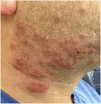
A 42-year-old man who works in an office presents with widespread grouped vesicles and pustules on an erythematous base on both sides of his beard (Figure 1). Most lesions are vesicular but some are purulent. They are itchy and painful.
The patient developed a single lesion initially. This was followed by additional lesions overnight, after shaving. He is systemically well but has some lymphadenopathy.
Differential diagnosis
Conditions to include in the differential diagnosis include the following.
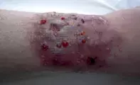
- Tinea barbae. This dermatophyte infection of the beard is relatively uncommon, but any unusual pustular eruption should raise suspicion of tinea. In hair-bearing areas, the lesions of tinea are characteristically dry and slightly erythematous. However, tinea can also be a highly inflammatory, vesiculopustular eruption with nodules, particularly when caused by a zoophilic organism (Figure 2). The lesions are often unilateral. Hair loss caused by broken hairs that fall out easily is a characteristic feature. For this patient, who developed more lesions on both sides of his beard after shaving, a tinea infection from a contaminated razor should be considered but it is unlikely because tinea infections tend to progress slowly and are not typically painful.
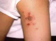
- Impetigo. This localised infection of the epidermis caused by Staphylococcus aureus or (occasionally) Streptococcus pyogenes can occur in patients of any age. Impetigo often affects the face, especially around the nose and mouth, but it can occur anywhere on the body. The lesions are characteristically erythematous and erosive, and vesicles or pustules typically evolve into crusted plaques that become enlarged and appear yellow, golden or brown in colour (Figure 3). There may be associated lymphadenopathy. It is possible for S. aureus to spread through shaving because it is usually passed from a carrier’s nose to normal skin. It would be highly unusual for the lesions to be painful.
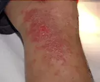
- Acute allergic contact dermatitis. This is a delayed hypersensitivity response to exogenous materials that contact the skin (e.g. a new shaving cream). The lesions are erythematous and intensely pruritic and tend to be plaque-like in nature. Severe lesions may have vesicles and even bullae that crust (Figure 4). Groups of vesicles, as seen in this patient, would be unlikely. The lesions tend to be itchy rather than painful.
- Sweet’s syndrome. This neutrophilic dermatosis can be idiopathic but it may also be induced by infection or drugs and it can be associated with an underlying malignancy. It is most common in middle-aged women. Typical findings include an eruption of painful plaques that are oedematous and erythematous. There may be evidence of pustules, bullae and, eventually, ulceration (Figure 5). There are usually multiple lesions. They can appear anywhere but are most often located on the arms, face and neck. Sweet’s syndrome would be unlikely in this patient, who was not unwell or febrile.
- Herpes infection of the beard. This is the correct diagnosis. Herpes infection of the beard (herpes sycosis) is most commonly caused by herpes simplex virus 1 (HSV1) and less commonly by HSV2. Reactivation of the latent virus can be triggered by stress, illness, trauma, fatigue or sun exposure, leading to acute and recurrent infections. HSV1 usually causes lesions around the lips and mouth, but in a minority of cases it can involve other areas of the face, such as the cheeks, chin and nose, as occurred in this case. The lesions tend to occur on an erythematous base and to be grouped, with multiple vesicles that may coalesce. Crusting is a typical feature of the lesions as they progress, and lesions are usually associated with significant pain and lymphadenopathy. Patients may also have symptoms of itch, fever, malaise, myalgia and joint pains; however, these are uncommon in patients with recurrent lesions, who tend to be systemically well. Lesions may be present inside the mouth, on the pharynx and hard and soft palates or on the perioral skin.
Investigations
HSV infections can be difficult to diagnose clinically when the presentation is atypical but it can be readily confirmed with laboratory testing. A viral swab of fluid taken from a deroofed vesicle should be taken and sent for polymerase chain reaction (PCR) testing and culture. A bacterial swab should also be taken to exclude any concurrent or secondary bacterial infection, which is not uncommon. If a fungal infection is suspected, it would be worthwhile to also take a skin scraping for culture and microscopy. For this patient, who most likely has a reactivation of a latent HSV infection (since the patient is systemically well), herpes serology is not useful and will only indicate that he has been exposed to HSV in the past.
Management
A HSV infection will usually resolve spontaneously over seven to 10 days. If the lesions are mild, small and uncomplicated then no treatment is necessary, especially if the area is kept clean and dry. However, for patients with herpes infection of the beard that is unsightly, painful and exposed to irritation from shaving, systemic treatment is appropriate. The patient described above was treated with oral valaciclovir (500 mg twice daily for seven days). There was a rapid improvement within 48 hours and the rash cleared by the end of treatment.
Topical antiviral therapy tends to have disappointing results for cutaneous HSV infections, particularly infections that are severe and widespread. Oral antiviral therapy with aciclovir, valaciclovir or famciclovir (off label) reduces the duration and the severity of the herpes infection, particularly when started promptly. Treatment of cutaneous herpes infection with oral antiviral medications is not covered by the PBS (unlike treatment of genital herpes infection). However, the cost of these medications has reduced recently as generic formulations have become available. Bacterial superinfection, if present, should be treated with appropriate oral antibiotics.
An attack of herpes may be a single event but there is always the potential for recurrence and patients should be warned about this possibility. For individuals who suffer recurrent attacks, long-term preventive antiviral therapy may be required, particularly if the attacks are severe. Alternatively, patients can be advised to keep oral antiviral medication on hand and to take it at the first sign of an impending attack, which can usually be identified by typical tingling sensations.

