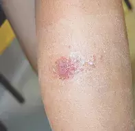
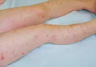
What’s the diagnosis?
A girl with an itchy vesicular rash


Case presentation
A 10-year-old girl presents with an intensely itchy vesicular rash on her lower abdomen that extends to her upper legs (Figure 1a). Some of the lesions are pustular. The rash was of acute onset, and occurred approximately one week after the sudden appearance of a lesion on her lower leg that is oedematous, crusted and exudative (Figure 1b). The girl is otherwise well and she has no history of dermatological problems.
Differential diagnoses
Conditions to consider among the differential diagnoses for a child of this age include the following.
- Scabies. Scabies should be considered in the differential diagnosis of any itchy papulovesicular rash. The presentation of scabies can be very variable and a high level of suspicion is necessary. Classically, scabies presents with erythematous papules, nodules, burrows in interdigital spaces and intense itch, particularly at night, which results in secondary excoriations. Vesicles and pustules, particularly on the palms and soles, can also be present in children. The diagnosis can be confirmed by direct visualisation of the scabies mite or its eggs using dermoscopy or direct microscopy of skin scrapings taken from burrows or nodules. Sometimes the best diagnostic test is observing response to appropriate treatment. The distribution of the rash in this case was not consistent with a provisional diagnosis of scabies.
- Folliculitis. This is a very common rash that presents as individual pustules associated with hair follicles (Figure 2). Each pustule is discrete on an erythematous base and the eruption is not typically itchy. The infection is usually caused by Staphylcoccus aureus, which can be isolated from a bacterial swab. For the case patient, folliculitis was unlikely to be the correct diagnosis because the rash was intensely itchy; in addition, bacterial swabs were taken and returned negative results.
- Molluscum contagiosum. This common viral rash is characterised by waxy umbilicated papules of varying sizes that are widely distributed over the limbs and torso. Each papule contains a viral core that can be expressed. Molluscum contagiosum is most common in children but can occur as a sexually transmitted infection in adults and it can be florid in immunosuppressed patients. The rash is generally asymptomatic but can be pruritic in children with a history of atopic dermatitis.
- Disseminated secondary eczema (autoeczematisation). This is the correct diagnosis. Also known as an ‘id reaction’, disseminated secondary eczema is an acute reaction pattern in response to certain triggers. It usually occurs days to weeks after an acute localised inflammatory skin disease. In children, disseminated secondary eczema is most often seen in association with tinea but it can also occur following allergic contact dermatitis and in adults is seen with venous stasis eczema or discoid eczema. The associated rash is acute, symmetrical and extremely itchy. It can present as poorly differentiated patches of eczema, vesicles or a morbilliform eruption. It is commonly distributed on the limbs and face but can occur on the trunk. The pathophysiology of disseminated secondary eczema is not well understood. The dissemination is thought to be haematological but it is not known what specific allergen is disseminated. The term ‘id reaction’ refers to an immunological process where there is an allergic reaction to an infection. In this case, it could also be called a ‘dermatophytid’.
Diagnosis
The diagnosis of disseminated secondary eczema is based on the identification of the primary lesion and exclusion of the differential diagnoses above. In the case patient, a fungal culture from scrapings from the lesion on her leg grew the dermatophyte Trichophyton rubrum and a swab of an abdominal lesion was negative for any bacterial growth. This dermatophyte, a primary human pathogen, usually causes tinea pedis and is unusual in children, who are much more commonly infected with dermatophytes acquired from animals. The origin of the fungus in this case was not apparent.
Management
The management of disseminated secondary eczema is twofold. The primary lesion needs to be treated aggressively while the id reaction requires anti-inflammatory treatment to resolve the intense itch. In this case, the patient was commenced on oral fluconazole 50 mg per day to treat the acute inflammatory tinea. The disseminated secondary eczema was managed with potent topical corticosteroid ointment (methylprednisolone aceponate). She recovered rapidly and all lesions resolved in two weeks. In cases where disseminated secondary eczema is more severe, crusted or exudative, wet wraps or oral prednisone may be required. Referral to a dermatologist is indicated if disseminated secondary eczema is suspected.

