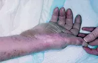
What’s the diagnosis?
Tense blisters with IgA deposits


Erythema multiforme and its more severe variants (Stevens–Johnson syndrome and toxic epidermal necrolysis) may be preceded by pharyngeal symptoms and subsequent multiple erythematous lesions that are often targetoid. Mucous membrane involvement is universal in severe cases and may be associated with widespread blisters that can appear burn-like (in toxic epidermal necrolysis). Skin biopsy demonstrates full thickness epidermal necrosis and lymphocytic inflammation. Skin immunofluorescence is usually negative. Drugs, including antibiotics and anticonvulsants, may induce this reaction.
Pseudoporphyria is associated with fragile acral skin and blisters that heal with scars. The sun-exposed dorsum of the hands and the face may be particularly involved. The blisters are not generalised, and mucous membranes are spared. Biopsy reveals a subepidermal blister without significant inflammation. Immunofluorescence may reveal a ‘coat sleeve’ deposition of immunoglobulin complement around vessels and at the basement membrane zone. These blisters may be induced by drugs, such as nonsteroidal anti-inflammatories (particularly naproxen) and tetracyclines, as well as by renal failure and sun beds.
Drug-induced pemphigus may present as erosive mouth lesions and flaccid, widespread, superficial blisters. Skin biopsy reveals intraepidermal clefting with loss of keratinocyte bonding (acantholysis). Skin immunofluorescence demonstrates intercellular deposits of immunoglobulin complement between keratinocytes. Drugs such as captopril, rifampicin and penicillamine may cause this reaction.
Linear IgA-induced bullous dermatosis is the correct diagnosis in this case. It is usually limited to the skin, particularly the flexures and acral areas, but mucous membrane lesions can develop. Skin biopsy revealed a subepidermal blister with neutrophils at the blister base. Linear deposits of IgA were found on immunofluorescence of frozen skin sections (Figure 2). The IgA linear deposits often occur in isolation. A wide range of drugs, including vancomycin, ciprofloxacin, captopril, lithium, furosemide, phenytoin and piroxicam, may induce this pattern. Treatment Discontinuation of the causative drug will usually result in resolution of the blisters over two to seven weeks. In severe cases, oral prednisone (25 to 50 mg daily in decreasing doses) or dapsone (100 mg per day) may be required.
A 63-year-old woman underwent cardiac surgery and developed a wound infection 10 days later. Culture grew methicillin-resistant Staphylococcus aureus. Vancomycin and ciprofloxacin were commenced. Prior medications included irbesartan and atenolol. Six days after commencing antibiotic therapy, widespread urticarial lesions appeared on the patient’s trunk and limbs. The lesions on the palms and soles rapidly developed into large blisters (Figure 1). There were no mucous membrane lesions.

