What’s the diagnosis?
A baby with a sudden blistering and erythematous skin eruption


Case presentation
A 10-month-old boy presents with an erythematous eruption that involves his whole skin but is accentuated in the flexures (Figure 1) as well as in the periocular and perioral skin. The eruption was of sudden onset, commencing 24 hours ago, and the child has become increasingly irritable, febrile and unwell. A number of superficial blisters and erosions are noted. His conjunctivae and oral mucosa are normal.
Differential diagnosis
Conditions to include in the differential diagnosis include the following.
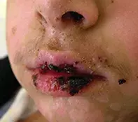
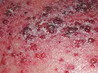
- Stevens–Johnson syndrome (SJS). This rare and serious mucocutaneous disease is characterised by conjunctivitis and epidermal and mucosal erosions, which often occur with severe stomatitis and scattered, subepidermal, blistering skin lesions (Figure 2). These lesions have a characteristic ‘target’ appearance, with blisters surrounded by erythema on a background of normal skin. There may be a febrile prodrome, with vomiting, diarrhoea, malaise and sore throat. In children, SJS is usually a reaction to infection with Mycoplasma pneumoniae or herpes simplex virus;1 less commonly, the syndrome is associated with drug reactions.2 Children with SJS are uncomfortable but on presentation they may not be unwell. SJS is unlikely in the presented case because of the complete lack of mucosal involvement and the generalised nature of the skin erythema.
- Viral skin eruption. Viral illnesses can manifest with varied cutaneous features and systemic signs. However, children with viral exanthemata are usually not severely unwell. The most common way for a viral skin eruption to present is as a blanching, erythematous, maculopapular eruption, but some patients have a blistering component (Figure 3). The rash is typically more severe on the arms, legs and face than on the trunk.
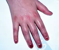
- Toxic shock syndrome. This illness has a sudden onset and is caused by infection with toxin-producing strains of Staphylococcus aureus or, less commonly, Streptococcus pyogenes. Children with toxic shock syndrome are acutely unwell and sometimes delirious. Patients have at least three involved organs that are damaged through poor tissue perfusion and direct damage from toxins. Children may have high fever, vomiting, diarrhoea, headache and myalgia. Hypotension is a characteristic feature, but may not be evident unless demonstrated by a postural drop. Early in the illness there is usually a diffuse erythematous exanthem that is most prominent in the flexural regions, but there is generally involvement of the conjunctivae and oral mucosa. The rash generally presents on the trunk before spreading symmetrically to the arms and legs. There is often erythema and oedema of the palms and soles (Figure 4). Vesicle and bullae formation (as occurred in the presented case) is not characteristic of toxic shock syndrome.
- Kawasaki disease. This is an acute febrile illness with associated vasculitis of the small and medium vessels with a predilection for the coronary arteries. It tends to affect infants and children under 5 years of age, and up to 25% of children with untreated Kawasaki disease develop coronary artery disease.3 The condition is usually characterised by a polymorphic eruption that is accentuated acrally. There can also be redness of the palms and soles with oedema (Figure 5). Kawasaki disease is unlikely in the presented case, as the patient is not displaying typical features such as fever for longer than five consecutive days, bilateral conjunctivitis, strawberry tongue and swelling of the cervical lymph nodes. Skin signs that are usually present in Kawasaki’s disease include erythema, bleeding, fissuring and crusting of the lips, oral cavity and pharyngeal mucosa. Vesicles and crusting, as occurred in this case, are uncommon.
- Drug reaction. An adverse reaction to a drug can cause a skin eruption but would usually not present as an asymmetrical blistering skin eruption. Drug reactions tend to result in erythematous, maculopapular or urticarial skin eruptions. Blistering is possible but rare and the rash is generally diffuse. The causative drug can usually be identified by taking a thorough history of medications (including over the counter medications and complementary and herbal preparations) used in the past two months. Children with drug eruptions are rarely severely unwell.
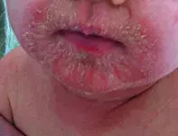
- Staphylococcal scalded skin syndrome. This is the correct diagnosis. Staphylococcal scalded skin syndrome (SSSS) is a blistering and then exfoliative skin eruption mediated by endotoxins A and B of S. aureus that cause superficial erosions. It is most common in infants and young children, especially those under 5 years of age, who have had a recent staphylococcal infection, which is not always evident and can be trivial. Conjunctivitis, otitis externa and minor skin infections have been implicated. The skin lesions of SSSS typically begin as erythematous patches that then blister, forming superficial bullae. These are easily de-roofed, leaving behind red and raw skin that is similar in appearance to a ‘scald’, which is often followed by crusting. The skin is fragile and may erode on pressure (Nikolsky’s sign). The skin eruption has a predilection for flexural, periorbital and perioral (Figure 6) regions, but it always spares the mucosa (unlike SJS). The toxin causes the skin to be very tender, which is why children with SSSS appear unwell and irritable.
Investigations
SSSS is diagnosed clinically and the source of infection should be determined if it is not evident immediately. Performing a septic screen is appropriate and should include blood culture and routine blood tests with full blood count, liver function testing, and electrolytes, urea and creatinine levels. Swabs should be taken from the skin lesions themselves and from the conjunctiva, nose, ears and axilla, and any recent areas of skin injury. It is not uncommon for a single skin swab to return negative results, so multiple swabs should be taken.
For a patient presenting with suspected SSSS, the most important diagnosis to exclude can be SJS; however, often a presentation of SSSS is so characteristic that an immediate diagnosis can be made. Differentiation between SSSS and SJS can be achieved without a biopsy by sending the roof of a blister for frozen section and histopathology frozen analysis. In SSSS only the stratum corneum will be present, whereas in SJS the frozen analysis will show the roof of the blister to be comprised of full-thickness necrotic epidermis.4
Management
Most children with SSSS require admission to hospital for analgesia, nursing care and treatment with either intravenous or oral flucloxacillin (50 mg/kg in four divided doses). As the patient recovers, the skin desquamates and treatment with emollients is indicated during this time. With adequate treatment, SSSS resolves over a period of 10 to 14 days and does not leave permanent scarring. The prognosis is very good. When the recovery is complete, the child and family members in their household should undergo a staphylococcal eradication protocol.
References
1. Wetter DA, Camilleri MJ. Clinical, etiologic, and histopathologic features of Stevens-Johnson syndrome during an 8-year period at Mayo Clinic. Mayo Clin Proc 2010; 85: 131-138.
2. Levi N, Bastuji-Garin S, Mockenhaupt M, et al. Medications as risk factors of Stevens-Johnson syndrome and toxic epidermal necrolysis in children: a pooled analysis. Pediatrics 2009; 123: e297-304.
3. Dominguez SR, Anderson MS, El-Adawy M, Glodé MP. Preventing coronary artery abnormalities: a need for earlier diagnosis and treatment of Kawasaki disease. Pediatr Infect Dis J 2012; 31: 1217-1220.
4. Handler MZ, Schwartz RA. Staphylococcal scalded skin syndrome: diagnosis and management in children and adults. J Eur Acad Dermatol Venereol 2014; 28: 1418-1423.
Infant and newborn care

